 Credit: The ROSAT Mission/Max-Planck-Institut für extraterrestrische Physik
Diagnosing the Heart of the Galaxy
"Tomography" is a technique in which a 3-dimensional picture is built up
slice-by-slice from 2-dimensional images. In medicine, positron emission
tomography (PET) or magnetic resonance imaging (MRI) is used to generate
3-d "X-rays" of the human body to provide vital information about many
forms of pathology. Tomography is also used by X-ray astronomers to
generate detailed images of matter in the universe. A tomographic
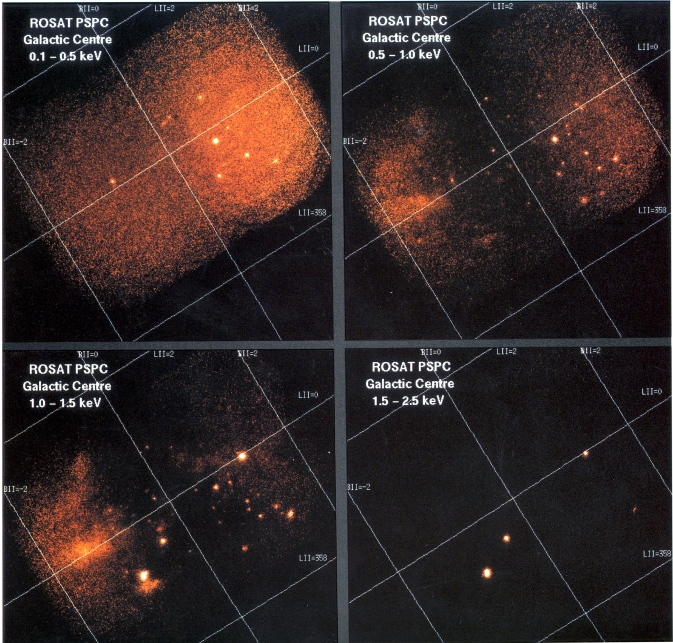
image of the center of the Milky Way Galaxy, obtained by the ROSAT X-ray observatory, is shown above.
The images represent X-rays of low (0.1-0.5 keV, upper left) to high
energies (1.5-2.5 keV, lower right). X-rays are absorbed by gas and dust
lying between us and the X-ray source; because high energy X-rays have
greater "penetrating power" than low energy X-rays, high energy X-rays can
pass through more of the interstellar medium than low energy X-rays. Since
the energy of the X-ray emission is a measure of the amount of interstellar
absorption, objects that emit only high energy X-ray emission are typically
more highly absorbed than objects which emit low-energy X-rays. Thus, by
measuring the x-ray energy being emitted, astronomers can develop a picture
of the mass distribution between us and the X-ray emitting source.
Last Week *
HEA Dictionary * Archive
* Search HEAPOW
* Education
Each week the HEASARC
brings you new, exciting and beautiful images from X-ray and Gamma ray
astronomy. Check back each week and be sure to check out the HEAPOW archive!
Page Author: Dr. Michael F.
Corcoran
Last modified April 17, 2001


