The high energy instrument PDS on-board the SAX X-ray astronomy satellite
F. Frontera1,2, E. Costa3, D. Dal Fiume1,
M. Feroci3, L. Nicastro1, M. Orlandini1, E. Palazzi1, and
G. Zavattini2
- 1 Istituto Tecnologie e Studio Radiazioni Extraterrestri, CNR, Via Gobetti, 101, 40129 Bologna, Italy
- 2 Dipartimento di Fisica, Universit`a di Ferrara, Via Paradiso, 11, 44100 Ferrara, Italy
- 3 Istituto di Astrofisica Spaziale, CNR, Via E. Fermi, 21, 00044 Frascati, Italy
Abstract
SAX/PDS experiment is one of four narrow
field instruments of the SAX payload, that also includes
two wide field cameras. The goal of PDS is to extend
the energy range of SAX to hard X-rays. The
operative energy range of PDS is 15 to 300 keV, where the
experiment can perform sensitive spectral and temporal
studies of celestial sources. The PDS detector is
composed of 4 actively shielded NaI(Tl)/CsI(Na) phoswich
scintillators with a total geometric area of 795 cm2 and
a field of view of 1.3o (FWHM). In this paper we
describe the experiment design and discuss its functional
performance and calibration data analysis system.
Key words: Instrumentation: detectors
1. Introduction
The SAX satellite is a joint program of the Italian Space
Agency (ASI) and the Netherlands Agency for Aerospace
Programs (NIVR) devoted to X-ray astronomical
observations in a broad energy band (0.1-300 keV). SAX is
scheduled to be launched in April 1996 into a circular
orbit at 600 km altitude with an inclination of about 3o.
The above inclination was chosen to get a high geomagnetic
cut-off with a very low modulation and to minimize the induced
radiation on the payload due to the
South Atlantic Anomaly (SAA). Overviews of the SAX
mission can be found elsewhere (Butler & Scarsi 1990;
Scarsi 1993; Piro et al. 1995). The satellite is
three-axis-stabilized with post-facto attitude reconstruction with 1
arcmin accuracy. Its expected lifetime is from 2 years
up to 4 years. The payload includes Narrow Field
Instruments (NFI) and Wide Field Cameras (WFC). The
NFIs are four Concentrators Spectrometers (C/S) with
3 units (MECS) operating in the 1-10 keV energy band
and 1 unit (LECS) operating in 0.1-10 keV, a High
Pressure Gas Scintillation Proportional Counter (HPGSPC)
operating in the 3-120 keV energy band and a Phoswich
Detection System (PDS) with four detection units
operating in the 15-300 keV energy band. Orthogonally with
respect to the NFIs there are two WFCs (field of view of
20o x 20o FWHM) which operate in the 2-30 keV energy
band with imaging capabilities (angular resolution of 5
arcmin). As a part of the PDS, there is a Gamma Ray
Burst Monitor (GRBM) with nominal energy band of 60
to 600 keV.
As can be seen, SAX covers an unprecedented wide
energy band from 0.1 to 300 keV. The hard X-ray energy
band (15-300 keV) covered by the PDS experiment is
now assuming key importance for high energy
astrophysics thanks to many exciting and unexpected
results obtained with gamma-ray astronomy experiments like
BATSE and OSSE aboard CGRO, SIGMA aboard the
GRANAT satellite and balloon experiments. In addition to
well known classes of strong hard X-ray emitters
(galactic X-ray pulsars, black hole candidates, Am
Hertype sources, AGN), other classes of X-ray sources, like
X-ray bursters, resulted to be strong hard X-ray
emitters during source states when the low energy emission is
relatively weak (Barret & Vedrenne 1994; Zhang et al.
1996). These sources were not expected to have hard
emission spectra. Soft gamma ray repeaters have been
discovered. Some of them exhibit parossistic phases of
activity, during which up to ~ 40 bursts per day
are detected (Kouveliotou et al. 1996, Lewin et al. 1996). Also
for extragalactic sources the hard X-ray sky was full
of unexpected results, e.g. for Seyfert 2 (Bassani et al.
1996).
With its high sensitivity, especially in the 20-100 keV
band, PDS will offer the opportunity to perform
observations of key importance in the hard X-ray band. Many
open questions in high energy astrophysics are expected
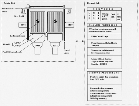 Fig. 1. A sketch of the major components of the PDS experiment.
to be clarified and solved with observations in this band.
Coordinated observations with GRO-BATSE and XTE
will give the opportunity to fully exploit the joint
capabilities of the three observatories that cover
this important energy band. Also coordinated observations with
on-ground and space observatories are extremely important
for identifying counterpart of new hard X-ray
sources and/or study the complex physics occurring in
them.
In this paper we will give a description of the PDS
and of its performances as derived from functional tests
and pre-flight calibrations. Descriptions of the PDS
design have already been reported (Frontera et al. 1991;
1992). Also reported are preliminary results obtained
from instrument functional tests (Frontera et al. 1996).
The GRBM expected performance has also been investigated
and results were reported (Pamini et al. 1990;
Alberghini et al. 1994).
2. Instrument description
A sketch of the major components of the PDS experiment
is shown in Fig. 1, and a summary of its main
features is given in Table 1.
The PDS consists of a square array of four independent
NaI(Tl)/CsI(Na) scintillation detectors. A system
Table 1. Main features of the PDS experiment
| PARAMETER | VALUE |
| Energy range | 15-300 keV |
| NaI(Tl) phoswich thickness | 3 mm |
| CsI(Na) phoswich thickness | 50 mm |
| Total area through collimator | 640 cm2 |
| Field of view (FWHM) | 1.3o hexagonal |
| Energy resolution at 60 keV | 15% (on the average) |
| Maximum time resolution | 16 micro sec
|
| Gain control accuracy | 0.25% |
| In-flight calibration accuracy | 0.1% |
| Minimum channel width | |
| of energy spectra | 0.3 keV |
| Maximum throughput | 4000 events s |
be independently rocked back and
forth to allow the simultaneous monitoring of source and
background. The gain of the detectors and of the
associated electronic chain is continuously monitored
and automatically controlled by using as sensor a
fixed calibration source (FCS) of Am 241 . The instrument can also be
periodically in-flight calibrated with a movable
calibration source (MCS). The AC lateral shields are also used
as gamma-ray burst monitor. Finally a particle monitor
(PM) provides information on high environmental
particle fluxes and triggers actions to prevent the damage
of the detectors photomultipliers (PMT). All events
detected in the PDS detector are processed by an Analog
Processor (AP), that includes all the analog electronics,
analog-to-digital converters (ADCs), AC logics, programmable
thresholds. Each detected event qualified as
a good one by the AP is time tagged and sent to a Digital
Processor (DP) for the data processing via a 40 bit
FIFO. The DP filters and formats the data that are finally
sent via Block Transfer Bus to the SAX On-Board
Data Handling (OBDH). The AP and DP constitute the
PDS Instrument Controller (IC).
In the following subsections the main elements of
PDS are described.
2.1. Detection plane
The detection plane of PDS is an array of four
scintillation detectors. Each of the four detectors is composed
by two crystals of NaI(Tl) and CsI(Na) optically coupled
and forming what is known as PHOSWICH (acronym
of PHOsphor sandWICH). The NaI(Tl) thickness is 3
mm and the CsI(Na) thickness is 50 mm.
The scintillation light produced in each phoswich
(PHW) is viewed, through a quartz light guide, by a
PMT EMI D-611. The NaI(Tl) acts as the X-ray
detector, while the CsI(Na) scintillator acts as an
active shield. The scintillation events (due to photons or
charged particles) detected by each phoswich can be
divided into three classes: those that deposit energy only in
the NaI crystal (NaI events or ``good events''); those that
deposit energy only in the CsI crystal (CsI events); those
that deposit energy in both crystals (mixed events). The
events of the first class are accepted, while the events of
the other two classes are rejected. The rejection is
performed using the different decay constants of the
scintillations produced in the NaI (about 0.25-6sec at room
temperature) and CsI (0.6-6sec and about 7-6sec for
CsI). The analysis electronics (see Section 2.7), via its
Pulse Shape Analyzer (PSA), is capable to discriminate
events of the classes described above. Figure 2 shows a
typical pulse shape spectrum in the 15-300 keV range.
It is characterized by two peaks, one (on the left) due
to NaI(Tl) events and the other due to CsI(Na) events.
The scale in abscissa is correlated with the decay time
of the scintillation pulses.
The selection of good events is performed through the
choice of lower and upper programmable PSA thresholds
in the Electronic Unit (see below).
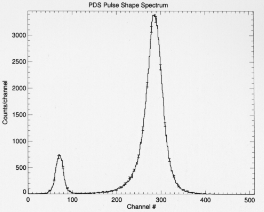 Fig. 2. Typical pulse shape spectrum in the 15-300 keV
energy range. Note the discrimination between NaI ``good''
events (left) and CsI events (right).
The phoswich configuration is well known to provide
a high detection efficiency and a very low background
level. In addition, taking into account that the accepted
photons lose energy in the NaI(Tl) alone, the energy
spectra provided by phoswiches are not affected by
biases as in single crystal detectors. The total geometric
detection area is 795 cm2. The other crystal parameters
are given in Table 1.
2.2. Collimator assembly
The collimator assembly consists of two X-ray
collimators made of Tantalum tubes with hexagonal section.
Inside each tube, a 4 cm bi-layer of Tin plus Copper
is inserted. The role of Tin, 100-6m thick, is to
attenuate K-fluorescence emission from the Tantalum
(Kalpha = 57.07 keV, Kbeta = 65.56 keV), while the Copper,
50-6m thick, was used to attenuate the K-fluorescence
emission from Tin (Kalpha = 25.1 keV, Kbeta = 28.5 keV).
This cell configuration is known as graded shielding. The
detector net area through collimators is 80% of the total
geometric area.
The collimators limit the detector field of view to
1.3o Full Width at Half Maximum (FWHM). Each of
the two collimators can be independently rocked back
and forth with respect to the neutral position directed
along the detector axis. Five symmetrical rocking steps
are provided on each side of the neutral position. The
maximum rocking angle is \Sigma 210 0 (3.5 degrees) and the
intermediate steps are \Sigma 30 0 , \Sigma 60 0 ,
\Sigma 90 0 , \Sigma 150 0 .
The standard observation strategy is to continuously
monitor background and source+background with both
collimators using a cyclic law: while one of the two
collimators points to the source the other one points to
a blank (non source-contaminated) sky field for background
measurement. At each cycle the two collimators
are swapped: the one pointing to the source is moved
to monitor background and viceversa. The default
collimator law samples alternatively two background fields
on the two opposite sides of the observed source at the
maximum rocking angle. Therefore the default full cycle
is ON /+210 0 (OFF) / ON /
w that rocks the collimators only on one side, or
that changes the rocking angle
to a value which excludes contamination, can be chosen.
It is also possible to move the collimators through all the
allowed positions. In this way one can derive the
angular response of the collimators to a given source and the
source position can be determined.
The dwell time (minimum 50 s) in each collimator
position can be chosen from ground.
A position transducer gives an independent high
accuracy (about 1 arcsec) monitoring of the collimator
position for each collimator. This monitoring is transmitted
to ground in the housekeeping (HK) data stream with a
sampling rate of 1 s.
2.3. Detector window
The X-rays entering through the telescope FOV cross
the top shield materials and the entrance window of the
phoswich crystals (kept under vacuum). The top shield
window is made of 1000 -6m BC-434 plastic scintillator
covered with aluminized Kapton (0.8 -6m Al plus 200 -6m
Kapton). The phoswich window is made of 1500 -6m Be,
890 -6m Silicone rubber, aluminized Kapton (0.1-6m Al
plus 76 -6m Kapton) and 100 -6m Teflon film. The
Silicone rubber was used to provide thermal and mechanical
insulation, the Teflon film was used as light reflective
material, while the aluminized Kapton was used as a barrier
between the Silicone rubber and the reflective material.
2.4. Anticoincidence shield system
A further reduction in the intrinsic background of PDS is
achieved using a five sides anticoincidence shield system.
This consists of the lateral and top shield assemblies,
both highly efficient in the rejection of events due to
charged particles. The CsI(Na) lateral shields are also
very efficient as active shields of photons coming from
the sides.
In the following we describe this subsystem and the
anticoincidence logics used to operate it.
2.4.1. Lateral shield assembly
The lateral shielding system is made of four optically
independent slabs of CsI(Na) scintillators 10 mm thick,
275 mm high and 402 mm wide. Each slab is made of two
pieces optically coupled together. Each slab is viewed,
through a quartz light pipe, by two 3 00 PMTs
(Hamamatsu R-2238) with independent high voltage power
supplies. In order to monitor the gain of each detector,
a light pulser made of an Am 241 source encapsulated in
a small NaI(Tl) scintillator is inserted into each slab to
produce pulses equivalent to photons with energy within
the range from 350 to 450 keV, depending on the particular
scintillator slab. The summed signals from each
slab are sent to a programmable threshold for the AC
logic. If the summed signal is above the AC threshold, a
veto signal is generated by the PDS analog processor.
2.4.2. Top shield assembly
The top shield assembly consists of a 1 mm thick plastic
scintillator (BC-434) viewed by four PMTs (Hamamatsu
R-1840) with independent power supplies. It is located
above the collimator assembly and covers the entire
telescope FOV. This shield was included in the PDS design
to reject the background events due to charged particles
entering the aperture of the instrument that deposit part
of their energy in the top shield. In particular we expect
that this shield is effective in rejecting events generated
by high energy electrons. These electrons are expected
to be produced by interactions of Cosmic Rays with the
satellite materials.
The summed signal from the four PMTs is sent to a
programmable analog threshold for the AC logic. If the
summed signal is above the AC threshold, a veto signal
is generated by the PDS analog processor.
2.4.3. Anticoincidence shield with other PHW units
Any event that is detected from one of the four PHW
units is compared with a programmable analog AC
threshold. If the signal is above this threshold, a logic
signal is set. A veto signal is generated by the PDS
analog processor if at least two units set this logic signal.
This happens when two or more simultaneous events are
detected from the PHW units above their A/C thresholds.
In this way we reject events that interact with one
of the NaI scintillators via Compton effect, leaving only
a fraction of their energy in it, while the photon with
the remaining energy is absorbed in other PHW unit(s).
2.4.4. AC logic and operative modes
The AC system acts as a logical OR among the AC veto
signals issued by the PDS Analog Processor. The
decision time of the AC logic is programmable with
5 equispaced steps between 2 and 10 -6s independently for each
�
F. Frontera et al.: The high energy instrument PDS
on-board the SAX X-ray astronomy satellite 5
of the AC signals described in the previous sections. The
AC logics are permanently activated with two operative
modes selectable from the ground: AC-ON in which the
vetoed events are not transmitted to ground; AC-OFF,
in which the vetoed events are flagged and transmitted
to ground. This last mode can be used only when PDS
is transmitting data in some Direct Modes (see Section
2.7). The AC of each PHW unit, of the lateral shields
and of the front shields can be separately programmed
to be ON or OFF.
2.5. Automatic gain control
The automatic gain control of the phoswich detectors
(AGC) is implemented as follows. At the center of the
detection plane there is a fixed calibration source (FCS).
It consists of an Am 241 radioactive source distributed
within a plastic scintillator (BC-400) viewed by a PMT
(Hamamatsu R 1635). Part of the 60 keV X-rays emitted
by this source impinges on the four phoswich detectors.
These events are tagged thanks to the 5.5 MeValpha
particles simultaneously emitted with them and detected by
the plastic scintillator.
The rate of 60 keV photons detected by each
phoswich as pure NaI events ranges from 3.6 to 4.4 s
PDS IC to detect gain variations and to perform a
continuous and automatic gain correction by acting on the
high voltage supplies of the phoswich PMTs. This
correction is operated using two prefixed contiguous channel
windows, with the separation channel corresponding to
the Am 241 60 keV peak position at nominal gain. Each
time a NaI event tagged as Am 241 event is inside the
left window or the right window, the HV of the
corresponding phoswich is changed by +97 mV or
to be used as an absolute calibrator, since the FCS
photons impinge only on a small
part of the phoswich detector surfaces, close to the
centre of the detection plane. Due to disuniformity in the
scintillation light collection across the crystal surface, a
bias in the detector gain should derive. Absolute gain
measurement can be performed using the in-flight
calibration system described in the next section.
2.6. In-flight calibration
The in-flight calibration of the PDS is performed by
means of two different devices: a Movable Calibration
System (MCS) and a light pulser.
2.6.1. Movable Calibration System
The movable calibrator MCS is made of a radioactive
source of Co 57 (main lines at 14.37, 122.06 and 136.47
keV) distributed along a wire of about 32 cm length,
and contained in a cylinder of lead that shields the
radioactive source but a small aperture directed toward
the crystal assembly. The MCS, upon an on-ground command,
performs a scan of the entire field of view of the
instrument above the collimators and top shield at constant
speed, uniformly irradiating all the PHWs. Given
the limited half life of the source (271 days), the
calibrator crossing speed can be varied in order to get the
needed statistical quality of the source data.
2.6.2. Light pulser
A light pulser (LED) is encapsulated in each phoswich
crystal. It allows to simulate a prefixed gamma equivalent
energy (GEE) lost in the phoswich. In this way
the gain of the phoswich PMTs and electronic chain can
be checked. The LED is mainly used for diagnostic purposes.
2.7. Electronic Unit
The electronic unit includes a power supply module and,
as discussed above (see section 2, the AP, which is
devoted to analog data processing and digital conversion,
and DP. This has in charge two tasks: the PDS data
management and the instrument control. The DP includes
two microprocessors (type 80C86): one (the Event
Processor, EP) to perform the first task and the other
(the Communication Processor, CP) to perform the second
task. Both processors make use of hybrid circuits.
The pulses from each phoswich unit with amplitude
in the band corresponding to the operative energy band
of PDS that pass the AC logic are pulse height and pulse
shape analyzed.
The AC logic allows to reject one or more of the
following types of events detected by the phoswich units:
- 1. Events simultaneous with events above the AC
threshold of the lateral shields;
- 2. Events simultaneous with events above the AC
threshold of the top shield;
- 3. Events simultaneous with events detected by other
phoswich units with energy above their AC threshold.
If one or more of the above coincided events are
accepted, they can be transmitted with the proper AC flag
in case of Direct Transmission Mode. By default the AC
logic rejects all the event types from 1 to 3 or any
combination of them.
The pulse shape analysis is performed using a constant
fraction/crossover PSA. For a detailed description
of the PSA and of its performance see Frontera et al.
1993.
The pulse height analysis is performed by a 12
bit successive approximation analog-to-digital converter
(ADC) with the sliding scale technique. This technique
was chosen to improve the average differential linearity
of the pulse height channels. The ADC conversion time
is 5 -6s.
The selection of the good events, corresponding to
energy losses only in the NaI(Tl) scintillator, is
performed through the setting of lower and upper pulse
shape digital thresholds. These thresholds are operated
by the EP.
The qualified events are formatted by the EP in
different transmission modes before being acquired by the
On-Board Data Handling (OBDH). The transmission
modes can be direct and indirect. Direct Modes (DM)
transmit information about each qualified event detected
by PDS. The information that can be transmitted to
ground is:
- energy: maximum 10 bits
- pulse shape: maximum 9 bits
- event time: maximum 16 bits
- unit identity: 2 bits
- AC flags: 3 bits (one for each AC type)
This maximum information can be compressed in two
ways: by masking some bits (the less significant bits) of
each field or by eliminating one or more fields. Fourteen
predefined compressions are available on board: the
maximum event size is 5 bytes (40 bits); the minimum event
size is 2 bytes (16 bits).
In Indirect Spectral Mode, the qualified events are
accumulated in energy spectra histograms, one for each
detection unit. For these integrated spectra the number
of energy channels (128 or 256), the maximum content of
each channel (8 or 16 bits), and the accumulation time
(5 choices, between 1 and 32 s) can be independently set.
The indirect spectral modes can be run in parallel with
temporal modes. These modes allow the transmission of
histograms with count rate time profiles in 1 to 4 energy
bands with time resolution down to 1 ms.
In addition to the information associated with good
events, other housekeeping (HK) information is provided
by the DP and transmitted. This includes pulse shape
spectra of the phoswich units in the entire energy band,
energy spectra of the FCS, various ratemeters, dead
time counters, status information and technological data
(high voltage and low voltage checks, temperature of
different instrument components, status of the MCS and
collimator motors). The HK spectra are summed on time
intervals of 128 s, the ratemeters are integrated over 1 s,
while the technological data are sampled and transmitted
with variable rate, from 16 to 64 s depending on the
data type.
2.8. Gamma-Ray Burst Monitor
The lateral shields of the PDS are also used as detectors
of gamma-ray bursts of cosmic origin (GRB). The
signals from each of the slabs in a programmable energy
band, nominally 60-600 keV (the low threshold can be
lowered down to about 20 keV), are sent to both a Pulse
Height Analyser and a GRBM logic unit, where the trigger
condition is checked. A GRB event is triggered when
at least two of the four lateral shields meet the trigger
condition. In this way we expect to reduce the rate of
false triggers caused by interactions of high energy
particles in the shields. If a GRB condition is verified, the
burst time profiles in the entire energy band from each of
the four detectors are stored, together with the absolute
time of the trigger occurrence. The integration time for
count accumulation will be 7.8 ms for 10 s preceeding
the trigger, 0.48 ms for 10 s immediately following the
trigger and 7.8 ms from 10 s to about 2 minutes after
the trigger time. Along with burst profiles, energy
spectra integrated over 128 s and ratemeters integrated over
1 s in the GRBM energy thresholds are continuously acquired.
Parameters of the trigger logic are programmable
from the ground. Details on GRBM and its performance
will be published in a separate paper in preparation.
2.9. Particle Monitor (PM)
A particle detector, made of BC-434 plastic scintillator
viewed by a PMT (R-1840), is mounted close to the PDS
instrument position in the satellite. The detector has a
cylindric-like shape with a diameter of 2 cm and a
thickness of 5 mm. It is covered on the top and sides with an
aluminum frame of 2 mm thickness. This configuration
provides an energy threshold that for electrons is about
1.2 MeV and for protons is 20 MeV. The PM is used
to switch off the high voltage supplies of all the PMTs,
excluding the PM PMT itself, when the environmental
charged particle count rate integrated over 32 s will
exceed a programmable threshold and to switch them on
again when this level is below another programmable
threshold.
3. Instrument Performance
The instrument performance was derived by means of
various tests performed at different levels. Functional
tests on the most critical items were performed at
component level (phoswich units, FCS, shield assembly,
collimators, PSA). Preliminary results of the functional tests
have already been reported (Frontera et al. 1996).
The instrument calibration was performed in successive
steps. First the Core Assembly (detection plane plus
FCS) was separately calibrated from the Shield Assembly
(top shield plus lateral shield plus MCS) and rocking
collimators. The rocking collimators were not integrated
in the detector in order to irradiate each phoswich
detection unit with almost uniform X-ray beams obtained
with radioactive sources at a distance of 1.8 m (see Section 3.2).
Then the core assembly was integrated with the
shield assembly with MCS source not mounted. A new
calibration of the detection plane was then performed
and compared with the previous one. Comparing the line
intensities measured with this calibration with the
corresponding ones measured before, we were capable to
get an experimental verification of the expected X-ray
transparency of the top shield.
Then the rocking collimators were integrated with
the detector. In this configuration the CsI(Na) slabs of
the lateral shields and the plastic scintillator of the top
shield were calibrated. Also the AC thresholds, the AC
decision time, the pulse height thresholds, the rise time
thresholds for selecting NaI events were calibrated and
set. The background level was also accurately measured.
Finally the MCS source was mounted in its frame and
more calibrations were performed and compared with
the previous ones. The detector contamination by MCS
source, its background behaviour as a function of
the collimator rocking angle and MCS count rate as a function
of the collimator offset were investigated.
AGC performance was investigated during the first
calibration campaign. After that, it was continuously
used during the subsequent calibration tests.
During all the calibration tests, the flight electronic
unit was used. Events from the phoswich units were all
acquired in direct mode, with the maximum information
associated to each event (see section 2.7). In addition all
the other available HK information were acquired. The
encoded data from the experiment were sent to a
simulator of the SAX OBDH and then to a mass memory.
In parallel, the encoded data from the OBDH simulator
were acquired by a PDS dedicated computer system (Dal
Fiume et al. 1994; 1995) for an on-line filing of the data,
quick look display, scientific analysis and archiving.
3.1. Monte Carlo tool
In order to derive the response function of the
PDS instrument, in addition to the calibration tests, a Monte
Carlo code, based on EGS4 (Nelson et al. 1985) was
developed. The geometry described in the simulation of
the detector included the top shield assembly, the four
phoswich units including the quartz light guides, and
the entrance window composition and the four Lateral
Shields. The X-ray interactions simulated by EGS4 code
include photoelectric absorption, Compton scattering,
K-fluorescence production and transport. All photons
were followed down to 5 keV. The transport of the
electrons produced in the photon interactions was
also included down to a minimum energy of 10 keV. Finally the
electronic analysis of the light pulses was calculated to
reproduce the pulse-shape analysis and energy spectra.
The pulse-shape distribution was reproduced assuming
a Poisson statistics of the light decay-time of the
scintillators and calculating the variance and covariance of
the electronic outputs. For the energy spectra the
measured calibration and resolution was used. We assumed
for the NaI(Tl) scintillation an exponential time
distribution with decay constant of 0.23 -6s, while for the
CsI(Na) we assumed the weighted sum of two exponential
functions with decay constants of 0.65 -6 s and 7 -6s,
respectively (Bleeker & Overtoom 1979). These values
are suitable to describe the scintillations at room temperature.
In this way we can compare the calibration spectra
measured with the expected ones as will be discussed in
section 3.3.3.
3.2. Radioactive sources for experiment calibration
The radioactive sources used for the experiment
calibration included Am 241 , Cd 109 , Co 57 , Ce 139 and Hg 203 ,
Na 22 , Sn 113 and Cs 137 . They were contained in proper
wells of Lead that acted as beam collimators. Each of
these sources was preliminary calibrated with their
collimator, by using an ORTEC HPGe detector with a
diameter of 25 mm, a sensitive thickness of 13 mm and
a X-ray entrance window of Beryllium 0.254 mm thick.
This preliminary calibration permitted to detect all the
gamma-ray lines (nuclear, fluorescence lines) with their
intensity emitted from the above sources and the presence of
very low level X-ray fluorescence from beam collimators.
3.3. Performance and calibration results
3.3.1. Detector window transparency
The whole transparency of the detection plane windows
(phoswich window and top shield window) as a function
of the photon energy was determined from the
knowledge of the material composition and thickness and it is
shown in Fig. 3. Also shown in this figure is the
computed transparency of the top shield window alone. The
test results were in complete agreement with the values
shown. The absorption coefficients were taken from the
EGS4 code data tables (Nelson et al. 1985).
3.3.2. Detector absorption efficiency
The X-ray absorption efficiency of the NaI(Tl) detectors
is defined as epsilon(E) = 1 -e-6(E)t , where 1-6 x (E) is the total
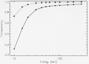 Fig. 3. Transparency of the detection plane window as a
function of the photon energy. Dashed line: top shield
window transparency. Full line: PHW composite window transparency.
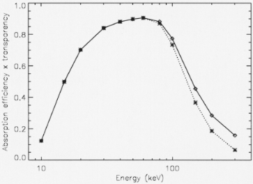 Fig. 4. X-ray absorption efficiency of the NaI(Tl) detection
units times the X-ray entrance window transparency. Dashed
line: only photoelectric absorption. Full line: total absorption.
absorption coefficient of the crystal and t its thickness.
The absorption efficiency times the transparency of the
PHW X-ray entrance window is shown in Fig. 4. The
thickness t was measured. The PDS detection efficiency
is also dependent on the pulse shape thresholds selected,
as it will appear from the next section.
3.3.3. Pulse shape discrimination
The pulse shape analysis is of key importance for the
experiment performance. The PSA discrimination
capability is very satisfactory as can be seen from Fig. 5, that
shows a pseudo image (pulse height channel PHA versus
PSA channel) of the Hg 203 radioactive source (nuclear
line at 279 keV, plus Tl K-fluorescence lines at ~ 85 and
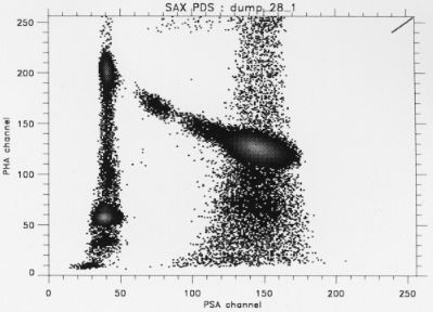 Fig. 5. A pseudo image of the Hg 203 radioactive
source (nuclear line at 279 keV), plus fluorescence X-ray lines at lower
energies due to Tl produced in the Hg decay, and to Lead of
the source collimator. It is apparent the separation of good
events due to NaI(Tl) scintillation (on the left) from events
due to CsI(Na) scintillation. It is also apparent a bridge
connecting the peaks, corresponding to energy losses in the
NaI(Tl) and CsI(Na), respectively. This is due to photons
that deposit part of their energy in NaI and part in CsI.
~ 72 keV). It is apparent the separation of the good
events due to NaI(Tl) scintillations (on the left) from
CsI(Na) scintillations (on the right) and mixed events
(in the middle).
Figure 6 shows the corresponding pseudo image obtained
with the Monte Carlo code. It is apparent the
similarity of the images apart from an apparent
deviation of the measured pulse shape distribution from that
obtained with the Monte Carlo code for low PHA channel
values that correspond to photon energies in the band
from 9-15 keV. This deviation is due to electronic noise.
It is also apparent in the simulated pseudo-image the
bridge connecting the two hot spots due to 279 keV
photons. It is due to Compton interactions of the 279
keV photons that deposit part of their energy in the NaI
crystal and part in the CsI crystal.
Figure 7 shows, for detection unit A, the PSA peak
centroid and width (2x FWHM) of NaI and CsI
crystals as a function of the PHA channel as derived from
background measurements. The measurements were
obtained at two different temperatures and the separation
is very good in both cases. It can be also seen that the
centroid position for both crystals is a function of crystal
temperature. For the NaI(Tl) we find results consistent
with those found in literature (Schweitzer & Ziehl 1983).
Good events are selected by choosing an electronic
digital window around ``pure'' NaI events. Given the
dependence of the pulse shape peaks on temperature and,
at a lower extent, on energy (see Fig. 7), the window
selection can affect the detection efficiency. A too narrow
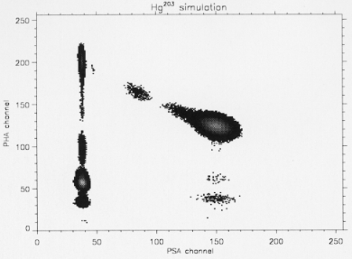 Fig. 6. A pseudo image obtained with a Monte Carlo
simulation corresponding to the Hg 203 radioactive source. It is
apparent the similarity with Fig. 5, apart from the lowest
energy (9-15 keV) photons pulse shape distribution. This
deviation is mainly due to electronic noise. Instead
is apparent in both the data and simulation images the bridge due
to Compton scattering.
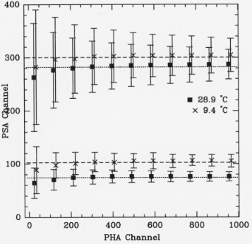 Fig. 7. Position of the pulse shape peaks of NaI and CsI
crystals as a function of the pulse height channel as
derived from background measurements. Squares (28.9o C) and
crosses (9.4o C) refer to measurement taken at different
temperatures. One error bar is equal to on FWHM of each PSA
peak.
window could decrease the detection efficiency, while a
broad window could include many Compton events and
thus introduce systematic errors in the spectral
reconstruction besides increasing the telescope background
level. Actually with our Monte Carlo code we can take
into account Compton interactions, single and multiple,
and thus obtain an unbiased spectral reconstruction.
A default PSA window (channels 1-70) was chosen
to derive the calibration results we report in this paper.
3.3.4. Energy to channel conversion
The nuclear and fluorescence lines emitted from the
available radioactive sources, that have an energy below
300 keV, were used to derive the relation between
line photon energy and pulse amplitude of the detected
line centroids. Figure 8 shows the net Co 57 spectrum as
detected by the phoswich unit A. It is apparent the 14
keV line (on the left) in the detected spectrum. In
order to derive the above relation, we fit the source spectra
with one or the sum of more Gaussian functions, depending
on line separation. For the X-ray fluorescence line
blends, we fit to the spectral data the sum of two
Gaussian functions due to the Kalpha and Kbeta lines, not resolved
in our detector. In order to limit the number of free
parameters, only the Kalpha line parameters (line centroid,
intensity and FWHM) were left free, while parameters of
the other lines in the ``blend'' were derived as functions
of the free parameters, making use of tabulated nuclear
spectroscopic data.
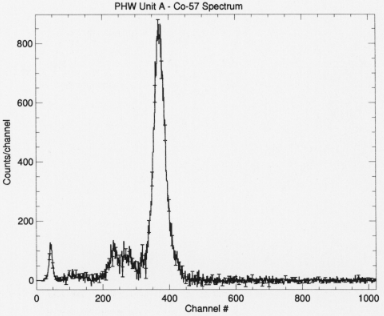 Fig. 8. Background subtracted energy spectrum of the Co 57
radioactive source used during calibrations as seen by Unit A.
The derived relation between line centroid channel
and photon energy is shown in Fig. 9 for each of the
four phoswich units. The AGC, that was activated during
all these tests, provided at 60 keV a gain equalization
of the phoswich units within 1.5%. A better equalization
(within 0.25%) was obtained during later tests at satellite
level. A clear feature of the energy/channel curves is
their almost similar behaviour up to 300 keV. This result
was obtained thanks to quality control of the NaI(Tl)
crystal production and to the fact that all the crystals
were cut from the same ingot.
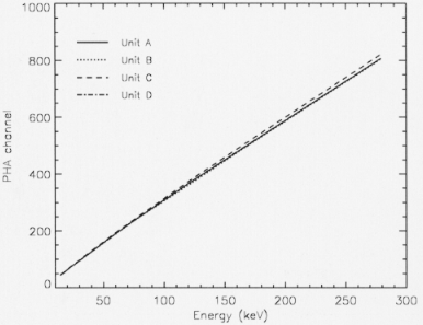 Fig. 9. The derived relation between PHA line centroid
channel and photon energy for the four PDS detection units.
The non-linearity curve of each NaI(Tl) crystal as a
function of photon energy, normalized to 279 keV Hg 203
line, is shown in Fig. 10. The curves are in agreement
with the expected behaviour of a crystal 3 mm thick
(see, e.g., Leo 1987). A detailed investigation of the
non-linearity of the NaI(Tl) around the Iodine K-edge
(33.170 keV) was not possible using only the available
radioactive sources. We plan to perform a thorough
investigation of the NaI(Tl) crystal non linearity with the
PDS flight spare, whose crystals were cut from the same
ingot as the flight model.
3.3.5. Energy resolution
The instrument energy resolution (R(E) = \DeltaE/E
where \DeltaE is the FWHM of the Gaussian that fits the
detected nuclear line of energy E) was derived by using
the same source spectra and results obtained to derive
the energy/channel relationship. The behaviour of the
energy resolution versus energy of one detection unit is
shown in Fig. 11. As can be seen, the data are not fit
with a unique power law R(E) / E
linear response up
to 300 keV. A broken power law can better fit the data.
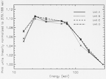 Fig. 10. The non-linearity curve of each NaI(Tl) crystal as a
function of photon energy, normalized to the 279 keV Hg 203
line.
Actually it is known (Sakai 1987) that the NaI(Tl)
scintillators show a deviation from the E
teraction position across
the crystal. This can also affect the energy resolution
for high energies where the fractional variation due to
Poisson statistics is lower. At 60 keV the average energy
resolution of the phoswich units is slightly better than
15%.
The dependence of the energy resolution on High
Voltage (HV) power supply of the phoswich PMTwas
investigated to determine the interval of HV within which
the phoswiches can be operated. Results have already
been reported (Frontera et al. 1996). In a range of \Sigma 200
V around the nominal HV (corresponding to 1170-1220
V depending on units) the energy resolution remains
constant.
3.3.6. MCS absolute calibration
Figure 12 shows the spectrum acquired from the instrument
during a passage of the MCS source across the
FOV. By comparing the parameters of the 122 keV line
of the MCS source with those obtained when a uniform
X-ray beam was incident on the same unit (see Fig. 8),
we found that the centroid positions and energy
resolutions are consistent each other. The typical
time duration of a calibration test is ~ 500 s with live time of the
MCS source above each phoswich of 200 s. The count
rate due to the 122 keV line was ~ 25 counts/s in March
1996. With this rate the peak centroid can be evaluated
with an accuracy of 0.1%.
Due to the decay of the MCS source, the calibration
time needed to get the same statistical quality of the
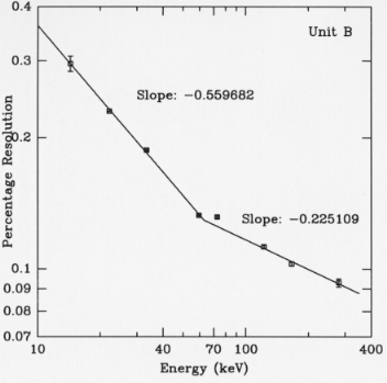 Fig. 11. The energy resolution of the detection unit B as a
function of energy. The slopes of two power laws are shown,
too. The break occurs at ~ 63 keV.
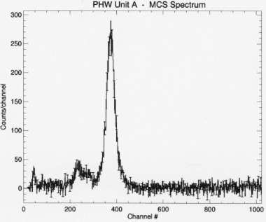 Fig. 12. Background subtracted energy spectrum of the Co 57
MCS source as seen by Unit A.
data will change with time. After two years the time
needed will be 6 times the initial one.
3.3.7. Effective area
The effective area of the detector units is limited at lower
energies by the overall composite window transparency
discussed above and at high energies by the NaI(Tl)
thickness. It also depends on energy resolution of the
detectors and, through it, on incident photon spectrum.
The effective area of the entire instrument as a function
of energy is shown in Fig. 13, for two different input
spectra (power law photon index equal to 1 and 2).
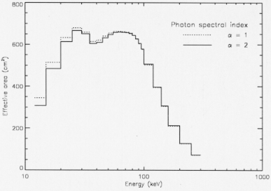 Fig. 13. PDS Effective area as a function of photon energy.
Two power law incident photon spectra of the form
3.4. Background counting rate
Great care was taken to minimize the PDS background
level. Indeed, the detector materials were selected with
low residual radioactivity. The phoswich technique
provides an efficient active shielding of the NaI(Tl) detector
over 2 pi solid angle, the lateral AC shielding
system provides a rejection of the unwanted photons and charged
particles, the AC top shield provides an efficient
rejection of charged particles, in particular electrons.
Three background contamination components were
identified: non tagged FCS source photons, residual
X-ray fluorescence due to rocking collimator materials and
contribution from gamma-ray lines of the MCS source.
The overall contribution from the first two components
gives rise to two main peaks in the background spectrum
at 60 keV and 30 keV with an integrated count rate of
about 0.3 counts/s per phoswich unit.
The third contamination component derives from
photons produced by Compton interactions with the
detector materials of weak high energy (?300 keV) lines of
the MCS source. The percentage integrated decay rate
due to these lines is 0.18%. For comparison, the
percentage decay rate of the 122 keV line is 85%.
This contamination component will decay along with the MCS
source decay. On the basis of the calibration tests
performed in March 1995, this component will contribute
with the following rates in September 1996,
corresponding to the beginning of the SAX operative phase: unit A:
0.45 counts/s; unit B: 1.1 counts/s; unit C: 1.1 counts/s;
unit D: 0.81 counts/s. As can be seen, different units
are affected in different way, depending on their position
with respect to the MCS source rest position.
The average background count rate/unit during
the system tests at the launch facility (March 1996)
was about 2.5 counts/s, corresponding to 4.4 x 10
in cm2 refers to the detector geometric area.
No significant background modulation with the rocking
collimator offset angle was detected. The upper limit
for this modulation, averaged on the entire energy band,
is ! 2% (3 oe). A detailed calibration of background
modulation is foreseen during the scientific verification phase.
4. Data Analysis
Concerning the PDS specific software, the following
modules will be included in the SAX data analysis software:
- gain and energy resolution versus energy, using calibration data;
- response matrix calculation (including tabulated
data coming from Monte Carlo simulations - see Section 3.1);
- background subtraction.
The third module will be tuned following the results of
the Scientific Verification phase.
Apart from this analysis software, a dedicated software
was developed at ITeSRE, based on a common
framework for SAX S/W (Chiappetti and Morini, 1991;
Maccarone et al. 1992), for the data acquisition and
analysis during ground calibrations (Dal Fiume et al.
1994; 1995). This software allows unpacking of raw data
from SAX telemetry, real time display of selected
housekeepings, analysis of calibration data. It can
transparently read data both in raw telemetry format and in
reformatted FOT format. The data display and analysis
section is based on IDL TM . A SQL database based
on Ingres TM was also configured to host the results of
the analysis of the PDS calibrations during all the
pre-operative and operative life of PDS. To this database,
we added external facilities to allow easy browsing of
the database. The choice of this SQL database was done
for the following reasons:
- high reliability due to the high quality of the DB
system;
- possibility to embed SQL calls in Fortran and C programs;
- capability to easily design and build GUI interfaces
using a native object oriented language.
This software configuration was designed to allow an
efficient control of PDS during all its life, with a stable
and coherent set of tools, allowing the PDS Scientific
Group to answer promptly to the needs of the scientific
community in terms of calibrations control, analysis and
results. A detailed description of the system will be published elsewhere.
Acknowledgements
The SAX program and thus the PDS experiment are
supported by the Italian Space Agency ASI.
Alenia Spazio (Torino), Laben SpA (Vimodrone, Milano),
Tecnol (Barberino del Mugello), Bicron (New Bury, USA)
and Officine Galileo (Firenze) with their industrial contracts
had a large part in the final PDS design as well as PDS make
up. We wish to thank many people who contributed to PDS
realization, in particular A. Basili, A. Emanuele, T.
Franceschini, G. Landini, E. Morelli, A. Rubini, S. Silvestri and C.M.
Zhang for their contribution to the preliminary PDS design
and M.N. Cinti who contributed to the analysis of the data
from functional tests. Finally warm thanks to R.C. Butler for
his continuous skilled and efficient supervision of the PDS final design and realization.
References
- Alberghini, F., Dal Fiume, D., Frontera, F., Pizzichini, G.
1994, AIP Conf. Proc. 307, 638
- Barret, D., Vedrenne, G., 1994, ApJS, 92, 505
- Bassani, L., Malaguti, G., Paciesas, W.S., Palumbo, G.G.C., Zhang, S.N. 1996, A&A in press
- Bleeker, J.A.M., Overtoom, J.M. 1979, NIM 167, 505
- Butler, R.C., Scarsi, L. 1990, SPIE 1344, 46
- Chiappetti, L., Morini, M. 1991, SAX/DAWG Report 15/91
- Dal Fiume, D., Frontera, F., Orlandini, M., Trifoglio, M. 1994 ASP Conf. Series 61, 395
- Dal Fiume, D., Frontera, F., Nicastro, L., Orlandini, M., Tri-foglio, M. 1995, ASP Conf. Series 77, 387
- Frontera, F. et al. 1991, Adv. in Space Res. 8, 281
- Frontera, F., et al. 1992, Il Nuovo Cimento 15C, 867
- Frontera, F., et al. 1993, IEEE Trans. on Nucl. Sci. NS-40, 899
- Frontera, F., et al. 1996, Il Nuovo Cimento C in press
- Kouveliotou, C., et al. 1996, Nat 379, 799
- Leo, W.R. 1987, Techniques for Nuclear and Particle Physics Experiments, Springer-Verlag Berlin Heidelberg
- Lewin, W.H.G., Rutledge, R.E., Kommers, J.M., van Paradijs, J., Kouveliotou, C. 1996, ApJ 462, L39
- Maccarone, M.C., et al. 1992, ASP Conf. Series 25, 151
- Nelson, W.P.R., Hirayana, H., and Rogers, D.W.O. 1985, SLAC-265
- Pamini, M., Natalucci, L., Dal Fiume, D., Frontera, F., Costa, E., Salvati, M. 1990, Il Nuovo Cimento 13C, 337
- Piro, L., Scarsi, L., Butler, R.C. 1995, SPIE 2517, 169
- Sakai, E. 1987, IEEE Trans. on Nucl. Sci. NS-34, 418
- Scarsi, L. 1993, A&AS 97, 371
- Schweitzer, J.S., Ziehl, H. 1983, IEEE Trans. on Nucl. Sci. NS-30, 380 Zhang, S.N., et al. 1996, A&A in press
